Comprehensive Study on Melasma: Etiology, Classification, and Therapeutic Interventions
Melasma, often referred to as the “mask of pregnancy,” is a common, chronic, and acquired hypermelanosis characterized by symmetric, brown-to-grayish-brown macules and patches. Primarily affecting the sun-exposed areas of the face, it is most prevalent in women of reproductive age and individuals with darker skin phototypes (Fitzpatrick scales III–V). While not physically painful or life-threatening, the psychological impact and social stigmatization associated with melasma are profound, often leading to decreased quality of life and emotional distress.
1. Introduction: The Complexity of Hyperpigmentation
Melasma is more than a simple cosmetic concern; it is a complex dermatological condition involving the over-activation of melanocytes—the cells responsible for pigment production. Unlike temporary tanning, melasma represents a functional derangement where the skin’s pigmentary system becomes hyper-responsive to internal and external stimuli.
The condition is notoriously recalcitrant, meaning it is difficult to treat and highly prone to relapse. Understanding melasma requires a deep dive into the interplay between genetics, hormonal fluctuations, and environmental stressors like ultraviolet (UV) radiation and high-energy visible (HEV) light.
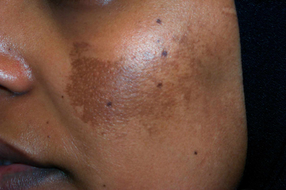
2. Symptoms and Clinical Presentation
The primary symptom of melasma is the appearance of discolored patches of skin. These patches are:
- Symmetrical: Usually appearing on both sides of the face in a similar pattern.
- Irregularly Shaped: The borders are often jagged or “geographic.”
- Variable in Color: Ranging from light tan to deep chocolate brown or even a slate-gray hue depending on the depth of the pigment.
- Asymptomatic: They do not itch, sting, or cause physical pain.
Common Locations:
- Centrofacial: Forehead, cheeks, upper lip, nose, and chin.
- Malar: Restricted to the cheeks and nose.
- Mandibular: Occurring along the jawline.
3. Types of Melasma
To treat melasma effectively, a dermatologist must determine the depth of the melanin. This is typically done using a Wood’s lamp (black light) examination.
| Type | Depth of Melanin | Appearance under Wood’s Lamp | Treatment Outlook |
| Epidermal | Superficial (top layer) | Becomes more distinct/darker | Most responsive to topical treatment. |
| Dermal | Deep (dermis layer) | Does not change or becomes less distinct | Difficult to treat; requires procedures. |
| Mixed | Both layers | Areas of both enhancement and fading | Requires a multi-faceted approach. |
4. Etiology and Triggers
The exact cause remains elusive, but several “master triggers” have been identified:
- UV and Visible Light: UV radiation induces lipid peroxidation in keratinocytes, releasing inflammatory mediators that stimulate melanocytes.
- Hormonal Influences: Pregnancy, oral contraceptives, and hormone replacement therapy are major drivers. The surge in estrogen and progesterone is believed to upregulate melanogenesis.
- Genetics: A family history is present in over 50% of cases.
- Inflammation: Heat and irritating skincare products can worsen the condition by triggering vascular responses.
5. Treatment Modalities
Treatment follows a “ladder” approach, starting with topical agents and escalating to procedural interventions.
A. Topical Gold Standards
- Hydroquinone (HQ): The primary tyrosinase inhibitor. It stops the conversion of DOPA to melanin. Usually prescribed at 2% to 4%.
- Triple Combination Cream (Kligman’s Formula): The most effective topical therapy, combining Hydroquinone, a Retinoid (to increase cell turnover), and a Corticosteroid (to reduce inflammation).
- Cysteamine: A newer, non-hydroquinone alternative that is gaining popularity for its safety profile.
- Tranexamic Acid (TXA): Used topically or orally, TXA inhibits the interaction between melanocytes and keratinocytes.
B. Chemical Peels
Peels like Glycolic acid or Salicylic acid help shed the pigmented upper layers. However, they must be used cautiously in darker skin tones to avoid post-inflammatory hyperpigmentation (PIH).
C. Energy-Based Devices (Lasers)
Lasers are reserved for resistant cases.
- Q-Switched Nd:YAG: Uses low energy to “shatter” pigment.
- Picosecond Lasers: Deliver ultra-short bursts of energy to treat dermal pigment with minimal heat damage.
2. Mechanistic Insights of Melasma
According to recent research, a number of factors, such as genetic predisposition, UV radiation, Photoaging, Hormonal factors etc. can cause or worsen the condition of Melasma, as shown in Fig. 1.

6. Aftercare and Maintenance
Melasma management is a marathon, not a sprint. The “maintenance phase” is where most patients fail.
- Strict Photoprotection: A broad-spectrum sunscreen with an SPF of 50+ is mandatory. It must contain physical blockers like Zinc Oxide or Titanium Dioxide, as these also protect against visible light.
- Tinted Sunscreen: Iron oxides found in tinted sunscreens are the only ingredients that effectively block HEV (blue light), which is known to worsen melasma.
- Heat Avoidance: Avoid saunas, hot yoga, and steam rooms, as heat can induce vasodilation and pigment production.
7. Conclusion
Melasma is a chronic condition that requires a lifelong commitment to skin protection. While complete “cures” are rare, significant clearance is achievable through a combination of medical-grade topicals, sun discipline, and professional procedures. Success depends heavily on the patient’s adherence to UV protection and the clinician’s ability to balance aggressive treatment with the prevention of irritation.