Wound and Scar Formation
A cutaneous scar represents a permanent, structural modification of the skin’s architecture following an inflammatory, traumatic, or surgical insult to the dermal tissue. When the protective epidermal barrier is breached and the injury extends deep into the papillary or reticular dermis, the skin’s natural repair mechanisms are instantly triggered to close the wound and prevent infection.
This repair process unfolds across four highly coordinated, overlapping biological phases:
- Hemostasis: Immediate vascular constriction and fibrin clot formation to halt bleeding.
- Inflammation: A rapid influx of neutrophils and macrophages to clear cellular debris and neutralize potential bacterial pathogens.
- Proliferation: The rapid migration of keratinocytes, angiogenesis to restore blood flow, and the deposition of a temporary extracellular matrix.
- Maturation (Remodeling): The progressive replacement of weak Type III collagen with stronger, organized Type I collagen fibers.
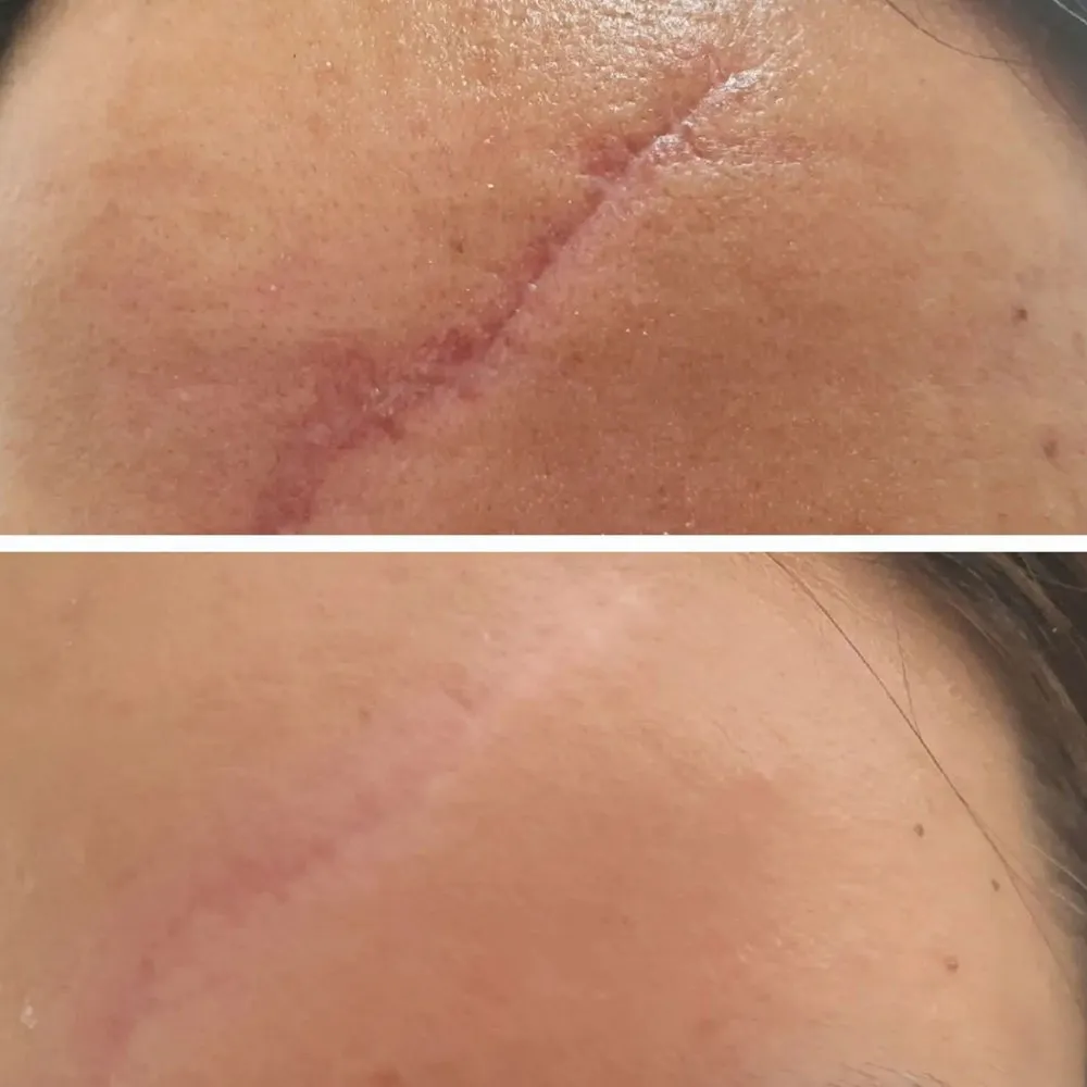
To address this issue effectively, we at Grazia Skin Clinics approach Scar Removal Treatment not as a superficial cover-up, but rather as an active intervention designed to disrupt and reprogram this biological remodeling phase. When the inflammatory phase of wound healing is either too short or prolonged, the delicate balance of collagen deposition and enzymatic degradation is disrupted.
- Under-production of Collagen: When the body fails to synthesize an adequate amount of extracellular matrix during the proliferative phase, the skin collapses inward over the site of injury. This structural deficit results in depressed, pitted indentations known as atrophic scars, which are highly common after severe inflammatory acne, varicella, or trauma.
- Over-production of Collagen: Conversely, when the inflammatory phase is prolonged, fibroblasts remain overactive for too long. They synthesize an excessive, disorganized tangle of collagen fibers that cannot be broken down by local enzymes. This excess tissue results in raised, firm, and often painful lesions known as hypertrophic scars or keloids.
Standard, over-the-counter scar creams and topical silicone sheets are fundamentally limited in their efficacy. While they can help retain moisture in the outermost layers of the skin, they are completely unable to penetrate deep into the dermis to break apart dense, fibrotic collagen bundles or stimulate fresh tissue remodeling. True structural correction requires advanced clinical treatments. By using precise medical lasers, targeted mechanical subcision, and specialized chemical reconstruction, we can safely clear away damaged tissue, release deep fibrotic ties, and reprogram your skin cells to build a smooth, beautifully uniform, and healthy skin surface.
The Clinical Typology of Scars
Because scars differ significantly in their structural depth, cellular makeup, and tissue density, clinicians require an accurate clinical diagnosis to design a successful treatment path. Scars fall into four primary pathological families:
1. Atrophic Scars (Depressed & Pitted)
Severe inflammatory acne vulgaris commonly triggers these scars, which exhibit a localized loss of dermal collagen, elastin, and subcutaneous fat.
- Icepick Scars: These narrow, deep, and sharply demarcated tracts extend vertically into the deep dermis or superficial subcutaneous tissue, appearing as if a sharp instrument punctured the skin.
- Boxcar Scars: Superficial or deep tissue loss causes these round or oval depressions, which feature steep, vertically defined borders that resemble chickenpox scars.
- Rolling Scars: These wide, shallow depressions feature gently sloping edges. Abnormal fibrous tethers that form in the subdermal layers cause this wave-like appearance by pulling the epidermis downward and creating an uneven skin texture.
2. Hypertrophic Scars (Raised & Confined)
These firm, raised, erythematous (red) lesions develop within the original boundaries of a surgical incision, burn, or traumatic wound. They typically emerge shortly after the injury, and excessive Type III collagen deposition drives their growth, though they may partially regress on their own over several years.
3. Keloid Scars (Invasive & Spreading)
Highly aggressive fibroproliferative lesions that extend well beyond the original boundaries of the initial skin injury. Driven by a genetic predisposition and sustained, low-grade inflammation, keloids continue to grow indefinitely over time. They present as firm, nodular, often painful or itchy masses that are rich in thick, highly disorganized collagen bundles.
4. Contracture Scars (Post-Burn)
These form when a large area of skin is lost, most commonly due to severe thermal burns. As the tissue heals, the surrounding margins contract aggressively to close the gap. Consequently, this tight contraction pulls the edges together, which can restrict normal joint movement and lead to chronic physical discomfort.
Comparison of Clinical Scar Classifications
| Scar Pathology | Depth of Tissue Damage | Principal Histological Feature | Primary Aesthetic Concern | Gold-Standard Clinical Intervention |
|---|---|---|---|---|
| Icepick (Atrophic) | Deep Dermis to Subcutis | High-density vertical fibrous tracts | Deep, narrow pitted holes | TCA CROSS, Fractional $CO_2$ Laser |
| Boxcar (Atrophic) | Mid to Deep Dermis | Focal loss of elastin and collagen matrix | Sharp, steep-walled craters | Erbium:YAG Ablative Resurfacing, RF Microneedling |
| Rolling (Atrophic) | Subcutaneous Fascia | Fibrotic bands anchoring epidermis to deep tissue | Wavy, uneven, shadow-casting skin | Surgical Subcision, Autologous PRP, Dermal Fillers |
| Hypertrophic | Papillary & Reticular Dermis | Excess, parallel-aligned collagen bundles | Raised, firm, red or purple cords | Cryotherapy, Intralesional Steroid/5-FU Injections |
| Keloidal | Deep Dermis to Perilesional Skin | Disorganized, whorled collagen collagenous masses | Invasive, painful, expanding nodules | Vascular Laser (PDL), Intralesional Therapy, Excision |
Benefits of Professional Scar Removal Treatment at Grazia Skin Clinic
Undergoing a customized, dermatologist-guided scar revision protocol offers profound structural, physiological, and emotional advantages over simple home remedies:
- Restoration of Structural Dermal Scaffolding: First and foremost, professional energy-based therapies safely penetrate the deep dermis to trigger a controlled healing response. Consequently, this process prompts the body to produce fresh, organized collagen and elastin fibers, filling out deep acne pits and smoothing out uneven contours.
- Release of Deep, Pulling Fibrotic Bands: In addition, manual subcision protocols physically sever the tough, deep-seated fibrous tethers that pull rolling scars downward. As a result, the skin’s surface instantly springs back to its natural height, instantly smoothing out uneven textures.
- Normalization of Tissue Pigmentation and Redness: Furthermore, targeted clinical treatments break up concentrated pools of brown melanin and seal overactive red blood vessels. Specifically, this vascular clearance eliminates the dark brown spots (Post-Inflammatory Hyperpigmentation) and persistent red marks (Post-Inflammatory Erythema) that commonly surround old scars.
- Safe Reduction of Raised, Hypertrophic Lesions: Finally, our medical-grade micro-injections and vascular lasers halt the overactive cell division feeding raised scars and keloids. Therefore, this targeted approach causes the thick tissue to safely flatten while simultaneously relieving chronic itching, burning, and physical discomfort.
Advanced Clinical Treatment Modalities
At Grazia Skin Clinics, we do not believe in a single, one-size-fits-all solution for scar tissue. To achieve flawless, smooth skin, our dermatologists design multi-step treatment protocols that combine several advanced clinical technologies.
1. High-Precision Ablative Laser Resurfacing
- Fractional Carbon Dioxide Laser : This state-of-the-art technology delivers matrixed micro-beams of high-energy laser light deep into the skin, creating thousands of microscopic treatment zones.
- Erbium:YAG Laser Resurfacing: Because the water in your skin cells rapidly absorbs this laser, our dermatologists can vaporize microscopically thin layers of scarred skin with extreme precision.
2. Deep Dermal Remodeling Protocols
- Radiofrequency Microneedling (RFMN): This procedure uses an array of ultra-fine, gold-plated insulated needles to bypass the outer epidermis and deliver precise radiofrequency heat energy directly into the deep dermis. In doing so, it creates controlled thermal injury zones at the exact depth of the scar tissue.
- Surgical Subcision (Nokor Needle Release): For wide, rolling scars, our surgeons gently insert a specialized tri-beveled Nokor needle parallel to the skin surface. Specifically, the surgeon moves the needle in a fan-like motion to physically slice through the tough, fibrous bands that anchor the scar to the deeper tissues.
3. Targeted Chemical Reconstruction
- TCA CROSS (Chemical Reconstruction of Skin Scars): For deep, narrow icepick scars, our specialists apply a high concentration of Trichloroacetic Acid with extreme precision directly to the bottom of the scar pit. By doing so, the high-strength acid induces a controlled chemical reaction that safely breaks down the vertical scar walls.
4. Intralesional Anti-Inflammatory Therapy
- Steroid and 5-Fluorouracil (5-FU) Micro-Injections: For raised hypertrophic scars and active keloids, we perform micro-injections of Triamcinolone Acetonide combined with the anti-metabolite 5-Fluorouracil directly into the scar tissue.
Meticulous Aftercare and Chronological Healing Timeline
The ultimate success of your scar removal treatment depends heavily on providing a clean, protected, and highly supportive environment at home while the treated skin cells actively regenerate.
- Apply Specialized Silicon-Based Gels: First and foremost, once the treated skin surface has fully closed, apply a thin layer of medical-grade silicone gel twice a day. By doing so, you create an invisible, protective barrier that locks in moisture and regulates collagen production, preventing the formation of irregular textures.
- Maintain Absolute Sun Protection: In addition, newly treated skin is exceptionally vulnerable to ultraviolet radiation. Therefore, applying a broad-spectrum physical zinc oxide sunscreen with an SPF of 50+ every 3 hours is vital to protect the healing epidermal cells from post-inflammatory dark spots.
- Keep the Dermal Barrier Deeply Hydrated: Furthermore, use a pure, non-comedogenic hyaluronic acid serum and ceramide-rich moisturizer twice a day. Specifically, this deep hydration supports the healing cells. Consequently, it accelerates the healing process while keeping the skin calm, soft, and comfortable.
- Avoid Physical Friction and Picking: Finally, allow any micro-crusts or peeling skin that form after laser or chemical treatments to flake off naturally on their own. Rather than helping, picking or scrubbing at these healing layers too early can disrupt the delicate rebuilding process and increase the risk of permanent scarring.
Conclusion: Reclaiming a Smooth, Restored Canvas
Scars can serve as permanent physical reminders of past injuries, skin struggles, or surgeries, but modern clinical dermatology ensures they do not have to dictate your skin’s future. You no longer have to settle for uneven textures, deep pitted shadows, or raised, uncomfortable marks.