The Comprehensive Guide to Skin Cancer: A Clinical Overview
1. Introduction: Understanding the Global Challenge
Skin cancer remains the most common form of cancer worldwide. As we move through 2026, clinical data indicates that while incidence rates are rising due to aging populations and environmental factors, the survival rates have never been higher thanks to early detection and precision medicine. At Grazia Skin Clinic, we believe that education is the first line of defense.
Skin cancer is the uncontrolled growth of abnormal skin cells. It occurs when unrepaired DNA damage to skin cells—most often caused by ultraviolet (UV) radiation from sunshine or tanning beds—triggers mutations that lead the skin cells to multiply rapidly and form malignant tumors.
2. The Primary Types of Skin Cancer
Not all skin cancers are the same. They are categorized based on the type of cells involved.
Basal Cell Carcinoma (BCC)
The most common form of skin cancer, BCCs are abnormal, uncontrolled growths or lesions that arise in the skin’s basal cells, which line the deepest layer of the epidermis.
- Characteristics: Usually develops on sun-exposed areas like the face, ears, and neck.
- Risk: They rarely spread (metastasize) beyond the original tumor site, but if left untreated, they can be highly disfiguring.
Squamous Cell Carcinoma (SCC)
The second most common form, SCC originates in the squamous cells that compose most of the skin’s upper layers.
- Characteristics: Often appears as scaly red patches, open sores, or elevated growths with a central depression.
- Risk: SCC has a higher likelihood of spreading to lymph nodes or distant organs than BCC if not caught early.
Melanoma
The most dangerous form of skin cancer. It develops in the melanocytes, the cells that produce pigment (melanin).
- Characteristics: Often resembles a mole or develops from one.
- Risk: Highly metastatic. If diagnosed and treated early, it is almost always curable, but if allowed to spread, it can be fatal.
Rare Forms
- Merkel Cell Carcinoma: A rare, aggressive skin cancer that typically appears as a flesh-colored or bluish-red nodule.
- Sebaceous Gland Carcinoma: An uncommon cancer originating in the oil glands.
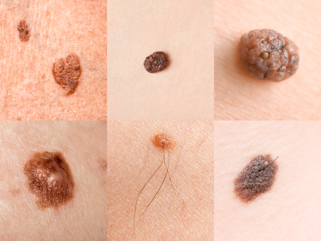
3. Symptoms and the ABCDE Rule
Early detection is vital. At Grazia Skin Clinic, we teach our patients the ABCDE Rule for identifying potential melanomas:
| Sign | Description |
| A – Asymmetry | One half of the mole does not match the other half. |
| B – Border | The edges are irregular, ragged, notched, or blurred. |
| C – Color | The color is not uniform and may include shades of brown, black, pink, or red. |
| D – Diameter | The spot is larger than 6mm (about the size of a pencil eraser). |
| E – Evolving | The mole is changing in size, shape, or color over time. |
General Warning Signs:
- A sore that does not heal within four weeks.
- A new spot or bump that looks different from surrounding moles (the “Ugly Duckling” sign).
- Persistent itching, tenderness, or pain in a specific area.
4. Causes and Risk Factors
While UV radiation is the primary culprit, several factors contribute to the development of skin cancer:
- UV Exposure: Intense, occasional exposure (causing sunburn) and cumulative lifetime exposure (sun tanning).
- Skin Type: Individuals with fair skin, light hair, and blue/green eyes have less melanin and higher vulnerability.
- Genetics: A family history of melanoma significantly increases risk.
- Immune Suppression: Those with weakened immune systems (e.g., transplant recipients) are at higher risk.
- Environmental Toxins: Exposure to substances like arsenic or coal tar.
5. Modern Treatment Options
At Grazia Skin Clinic, treatment is tailored to the cancer type, size, and location.
Surgical Interventions
- Mohs Micrographic Surgery: The gold standard for BCC and SCC. The surgeon removes thin layers of skin and examines them under a microscope immediately until no cancer cells remain. This spares the maximum amount of healthy tissue.
- Excisional Surgery: The tumor and a margin of healthy skin are cut out.
Non-Surgical & Advanced Therapies
- Cryotherapy: Freezing the cancer cells with liquid nitrogen.
- Immunotherapy: Boosting the body’s immune system to recognize and destroy cancer cells (commonly used for advanced melanoma).
- Targeted Therapy: Drugs that “target” specific molecules inside cancer cells (e.g., BRAF inhibitors).
- Photodynamic Therapy (PDT): Using a light-sensitizing agent and special light to kill abnormal cells.
6. Clinical Aftercare and Long-term Management
Recovery doesn’t end when the lesion is removed. Proper aftercare ensures minimal scarring and prevents recurrence.
Immediate Wound Care
- Keep it Clean: Wash the area 1–2 times daily with mild soap and water as directed.
- Moisturize: Apply a thin layer of petroleum jelly (Vaseline) to keep the wound moist, which promotes faster healing and less scabbing.
- Protection: Keep the surgical site covered with a sterile bandage for the first 24–48 hours.
Scar Management
Once the wound has closed, we recommend:
- Silicone Gel/Sheets: To flatten and soften the scar.
- Sun Block: Scars are highly sensitive to UV; darkening (hyperpigmentation) can be permanent if not protected.
The “Grazia” Prevention Protocol
- Annual Professional Exams: A full-body check by a dermatologist.
- Monthly Self-Exams: Use mirrors to check hard-to-see areas like the scalp and back.
- Broad-Spectrum SPF 30+: Apply daily, even on cloudy days.
- Protective Clothing: Utilize UPF-rated fabrics and wide-brimmed hats.
Note to Patients: Skin cancer is highly treatable when caught early. If you notice a spot that is “new, changing, or unusual,” contact Grazia Skin Clinic immediately for a professional evaluation. Your skin is your body’s largest organ—let’s protect it together.